Conditions
Retinal tears & Retinal detachment
While retinal tears and retinal detachment are painless conditions, they may lead to visual impairment or vision loss if left undiagnosed and untreated.
What are retinal tears & retinal detachment?
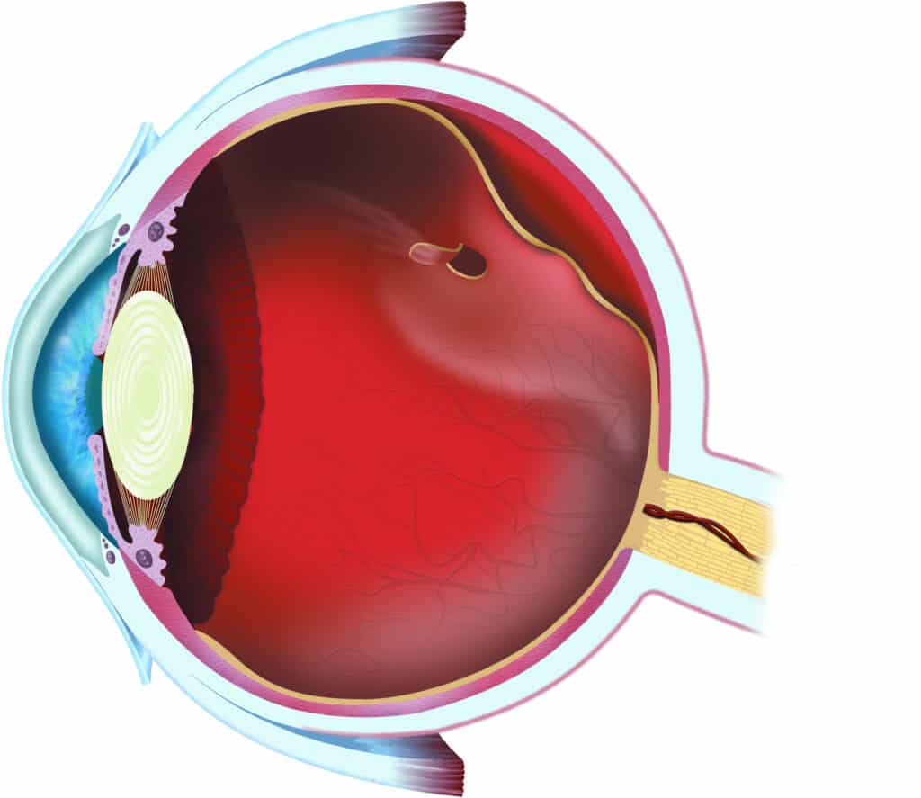
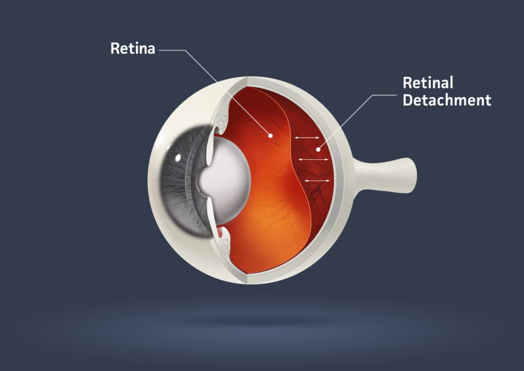
Retinal tears and retinal detachment are two types of vitreous-related degenerative conditions that affect the vitreous (the gel-like substance that fills the back of your eye) and result in the retina separating from the back of the eye.
Your retina is a thin layer made up of photoreceptors; cells that transmit light signals to your brain so you can see.
When the retina is torn or detaches from the back of your eye, your sight is reduced and you could experience complete vision loss if this retinal detachment is not treated by an experienced ophthalmologist.
Vitreous-related conditions include the following:
- Retinal tear
- Retinal detachment
- Retinoschisis
- Lattice degeneration
- Macular traction

Retinal tear
A retinal tear is a rip in the retinal layer when the vitreous pulls on the retina (often, but not always, caused by a posterior vitreous detachment or PVD).
A related condition is a retinal or macular hole which occurs when the retina becomes too thin and the photoreceptors degenerate or die, reducing the retina’s ability to transmit light signals to your brain.
Both retinal tears and retinal holes can lead to proliferative vitreoretinal membranes (PVR) and partial or complete retinal detachment.
When a retinal tear occurs, fibrotic cells become fibrous tissue and attach themselves to the retina, distorting it and impairing its ability to transmit light signals effectively.
This is known as subretinal fibrosis.
In order to treat and prevent retinal detachment, a retinal surgeon may elect to treat the subretinal fibrosis by removing PVR membranes from the retina.
Retinal detachment
Retinal detachment occurs when a retinal tear or hole is left untreated.
Retinal detachment can be described as chronic or macular, and may lead to post-detachment epiretinal membrane complex.
Chronic retinal detachment develops when a retinal tear or hole is untreated for a long time.
Once chronic retinal detachment has been identified, your ophthalmologist will typically treat this condition with intraocular injections or a surgical procedure to prevent permanent vision loss.
Macular detachment is less severe than chronic detachment and develops when the macula–the part of your eye responsible for central vision–detaches from the back of your eye.
Both chronic and macular detachment can result in subretinal fibrosis.
Retinoschisis
Retinoschisis describes a form of retinal detachment where the different layers of the retina split from each other causing partial or complete vision loss.
Macular traction
Macular traction is a form of partial posterior vitreous detachment or PVD that affects the macula.
Lattice degeneration
Lattice degeneration refers to a long-term, chronic thinning of the retina that may lead to a partial or complete retinal detachment.

What causes retinal tears & retinal detachment?
Retinal tears and detachment are caused by one or more of the following conditions:
- An eye injury
- A blow to the head
- Certain eye diseases
- Eye surgeries
- Diabetes
- Myopia
- Age-related thinning of the retina
- A build-up of fluid under the retina
- Posterior vitreous detachment (PVD)
Retinal tears & retinal detachment symptoms
Retinal tears and detachment don’t typically cause you pain; however, if your retina is partially or completely detached, you may experience the following symptoms:
- Seeing flashing lights at the edge of your vision
- Specks, lines, or spots (floaters)
- Decreased peripheral or central vision
- Blurry or veiled vision


Retinal tears & retinal detachment diagnosis
Retinal tears and detachment are diagnosed using a dilated eye exam that includes:
- A visual acuity test
- A pupil response test
- A tonometry test
Your ophthalmologist uses these tests to evaluate:
- How clearly you can see from various distances.
- How good your peripheral vision is.
- How well your pupils react to light.
- The pressure inside your eye (intraocular pressure or IOP).
Your eye doctor may also assess the condition of the blood vessels inside your eye with the use of an ultrasound exam.

Retinal tears & retinal detachment treatment
Retinal tears and detachment usually require surgical procedures to achieve two immediate objectives:
- Reattach the retina
- Prevent further vision loss
Retinal detachment surgery includes the following:
- Panretinal photocoagulation (PRP)
- Laser retinopexy
- Pneumatic retinopexy
- Scleral buckle
- Vitrectomy
- Silicone oil
- Proliferative vitreoretinopathy (PVR) stripping
The sooner after diagnosis the treatment occurs, the more successful the retinal detachment surgery will be.
Why choose Vision Pros to treat your retinal tears and retinal detachment?
- Experienced ophthalmology team with expertise in all areas of ophthalmology, optometry, and general eye care.
- Rapid access and referral to world-class comprehensive screening and cutting-edge treatment in a state-of-the-art ophthalmology clinic.
- Holistic, personal, and patient-centric approach to every aspect of eye care.
Book an appointment for your comprehensive eye examination today
- https://www.nei.nih.gov/learn-about-eye-health/healthy-vision/get-dilated-eye-exam
- https://www.nei.nih.gov/learn-about-eye-health/eye-conditions-and-diseases/retinal-detachment/laser-surgery-and-freeze-treatment-retinal-tears
- https://www.nei.nih.gov/learn-about-eye-health/eye-conditions-and-diseases/retinal-detachment/surgery-retinal-detachment
- https://my.clevelandclinic.org/health/diseases/10705-retinal-detachment
- https://www.medret.ca/retina-laser-treatments/
- https://www.medret.ca/vitreous-disease/
- https://www.nei.nih.gov/learn-about-eye-health/healthy-vision/get-dilated-eye-exam
- https://opto.ca/eye-health-library/retinal-detachment
- https://occeyecare.ca/RETINAL_DETACHMENT
- https://calgaryretina.ca/retinal-conditions/retinal-tear/index.html
- https://myhealth.alberta.ca/Health/Pages/conditions.aspx?hwid=hw187829